Mohs Surgery
What Is Mohs Surgery?
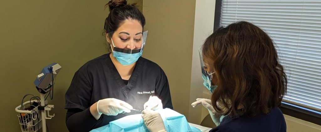
Mohs micrographic surgery, which is performed in our office using local anesthesia, is a state-of-the-art treatment for skin cancer in which the physician serves as a surgeon, pathologist, and reconstructive surgeon. It yields the highest cure rate of all skin cancer treatments, with cure rates approaching 99% for tumors that have not previously been treated. The procedure relies on the precision and accuracy of examining tissue under a microscope to trace and ensure removal of the skin cancer, which may have roots that extend beyond what is visible to the naked eye. In addition, the removal of healthy skin is minimized, resulting in the smallest possible surgical defect and thereby decreasing the potential for scarring.
Mohs micrographic surgery was developed by Frederic E. Mohs, M.D. in 1936 at the University of Wisconsin. In its original form, described as “chemosurgery”, the technique utilized a 20% zinc chloride fixative paste that was applied directly to the skin of the patient for fixation of the tissue. Subsequently, the involved skin was surgically removed by serial excision with microscopic control of the tissue margins.
Specialties
- Aesthetician Services
- Agnes RF
- Blue Light & Levulan
- Botox Cosmetic
- Cellulaze
- Chemical Peels
- Clear + Brilliant Laser
- CoolSclupting
- CoolTone
- Cosmopen
- DAXXIFY
- Dermal Fillers
- DermaSweep
- DiamondGlow
- Elite Laser
- FotoFacial
- Fraxel Dual Laser
- FX Laser Resurfacing
- Hair Restoration
- HydraFacial
- KeraLase
- Kybella
- Latisse for Lashes
- LaseMD Skin Resurfacing
- Laser Hair Removal
- MiraDry
- PicoSure Laser
- Scarlet SRF
- Scar Revision
- Sclerotherapy
- Skin Cancer Reconstruction
- SmartLipo
- SofTap
- Tattoo Removal
- Ultherapy
- UltraClear
Removal of tissue was performed in layers and color coded with dyes in order to orient specimens to the patient. Dr. Mohs created a unique technique of color-coding excised specimens and developed a mapping process to accurately identify the location of remaining cancerous cells. This original “chemosurgery” technique, which is no longer performed, was very painful and sometimes took days to complete.
The surgical procedure has been extensively refined over the last seven decades, but it still relies on the fundamental principles of color-coded mapping of excised specimens and their thorough microscopic examination. Surgeons now excise the tumor in layers and examine the fresh tissue immediately. This reduces the normal treatment time to one visit and allows for immediate reconstruction of the wound.
In 1967, the American College of Mohs Micrographic Surgery and Cutaneous Oncology was formed to recognize surgeons who have completed specific, certified training in the Mohs technique. The College also functions as a regulatory and certification body for over 60 Mohs fellowship training programs and provides a source of continuing education for more than 700 practitioners of Mohs Micrographic Surgery.
Mohs Surgery Information
Mohs Micrographic Surgery is primarily used to treat basal and squamous cell carcinomas. Mohs Surgery is indicated when:
- The borders of the cancer are poorly defined
- The cancer is large
- The cancer has recurred after a prior treatment
- The cancer has previously been incompletely removed
- The cancer is in an area where preservation of healthy tissue is paramount to achieve the best possible cosmetic and functional reconstructive outcome, such as the nose, ears, eyelids and lips
- The cancer has grown rapidly
- The cancer has aggressive microscopic features, such as morpheic, infiltrating, or micronodular
- The cancer tracks along nerves (perineural invasion)
Mohs Micrographic Surgery provides the highest possible cure rate of all skin cancer treatments. The cure rate for basal cell carcinomas that have not been previously treated exceeds 99% and the cure rate for previously untreated squamous cell carcinomas is 97%. The cure rates for recurrent basal cell carcinomas and squamous cell carcinomas are 97% and 90%, respectively.
The high cure rate provided by Mohs surgery is due to the unique and highly specialized tissue processing which occurs after the skin cancer has been removed from the patient. With Mohs surgery, horizontal sections are cut from the specimen. This is analogous to using a cheese slicer to remove a thin layer from the edges and base of the specimen with a single cut. This allows 100% of the peripheral and deep margins to be assessed so that any areas where skin cancer cells extend beyond the wound edges will be detected.
In contrast, other non-Mohs types of tissue processing used in skin cancer treatment involve vertical sectioning, in which the specimen is cut like a loaf of bread and the edges of the slices “sampled.” With this technique, less than 1% of the true surgical margins are assessed, which may result in a failure to detect areas of cancer that have extended past the surgical margins.
Thus, it is the way in which the tissue is processed that makes the Mohs procedure unique and provides the highest possible cure rate in treating skin cancer.
The Mohs Micrographic Surgery procedure relies on a specific sequence of surgery and pathological investigation:
- The surgeon identifies the visible skin cancer.
- The skin cancer and surrounding skin are anesthetized using local anesthesia, which is injected using a small needle.
- The visible skin cancer and a narrow margin of healthy-appearing skin are surgically removed with a scalpel and marked with reference points on the patient.
- An anatomic map of the removed tissue is created, which will guide the surgeon to the precise location of any residual cancer cells.
- The tissue specimens are labeled with dyes that allow the surgeon to reference the tissue seen on the microscope slides directly back to the specific location on the patient.
- The pathology technician processes the tissue specimens in the office lab and procedures frozen section slides.
- The slides are examined under a microscopic by the Mohs surgeon.
- If any of the microscopic sections contain tumor (i.e., skin cancer cells), the map guides the surgeon to the precise location where tumor roots remain.
- An additional layer of tissue is removed in the location of the roots, and the tissue is processed and examined as previously described.
- The tumor resection follows the same step-by-step process until all microscopic sections are clear from cancerous cells.
- At this time, the surgeon will discuss options for reconstructing the surgical defect with the patient.
The best method for reconstructing the surgical wound is determined after the cancer has been completely removed and the extent of the surgical defect has been determined. The primary considerations are preservation of function and maximizing the aesthetic outcome. Small wounds may be allowed to heal on their own, or the wound may be closed with stitches. Facial wounds often require reconstruction with a skin flap, or sometimes a skin graft. Reconstructing a wound with a skin flap involves utilizing lax skin that is adjacent to the wound, whereas a skin graft is performed by removing skin from another area of the body. The benefits and risks of each option will be discussed with you at the time of your surgery.
Surgery FAQs
The length of time required to complete a Mohs surgery case is variable, and is based on the involvement of the tumor and the complexity of the surgical reconstruction. As a general rule, each stage of Mohs will require approximately one hour for tissue processing to be completed. If the tumor persists at the margins, an additional stage will be performed, which will add an additional hour to the procedure. Additional stages, if necessary, are performed until all of the cancer has been removed. The surgical reconstruction will then take an hour or more to complete. Given the highly variable time requirements to complete the Mohs procedure, we ask that all patients anticipate being in the office all day and plan their schedules accordingly. Much of the day involves waiting in the office’s reception area or the examination room while the tissue samples are being processed.
As the Mohs procedure is performed under local anesthesia, patients are encouraged to have a normal breakfast on the morning of their procedure. Patients are asked to avoid caffeine on the day of their surgery, however, as caffeine, in combination with the epinephrine that is typically used in the local anesthetic, may result in over-stimulation.
Many patients drive themselves to their Mohs surgery appointment. As the procedure is performed using local anesthesia, there is no contraindication for driving post-operatively. However, patients who receive oral pain medications or anti-anxiety medications during the procedure are required to have a driver. In addition, patients with cancers around the eyes or nose may have swelling post-operatively, which may partially obstruct one’s vision and necessitate a driver.
The Mohs procedure is performed using local anesthesia. Prior to administrating the local anesthesia, which is injected with a small needle, a topical anesthetic is often used to minimize the discomfort of the injection. Additional local anesthetic is administered throughout the day, if necessary, to minimize discomfort. Oral medications are also sometimes used to control pain and minimize anxiety, if necessary.
A successful surgical outcome relies on the skill of the surgeon, appropriate post-operative care by the patient, and the ability of the body to heal. The Mohs procedure minimizes the removal of healthy skin, resulting in the smallest possible surgical defect. This helps to decrease the risk of scarring. Your Mohs surgeon will choose the most appropriate reconstruction option to achieve the best possible aesthetic outcome. Post-operative wound care instructions are provided, which patients must follow closely to promote healing. The majority of patients heal with an imperceptible scar. Some wounds, however, may take several months to completely settle. A small percentage of patients will require a touch-up surgery or laser procedure to achieve the highest possible aesthetic outcome.
There is typically some degree of bruising and swelling after the Mohs procedure, particularly for surgeries around the eyes or lips. These changes are most pronounced during the first one to two days and may take up to a week or more to resolve. A larger bandage is typically required for the first one to two days, at which time a smaller bandage is placed. Many patients return to work the day after the procedure, if they are comfortable going to work with a larger bandage and some degree of bruising and swelling.
Sutures on the face and neck are removed between days 5 and 7 post-operatively. Sutures on the scalp, torso, and extremities are removed between 12 and 14 days post-operatively.
